Myelinated Neuron Anatomy is a photograph by Science Photo Library which was uploaded on June 28th, 2016.
Myelinated Neuron Anatomy
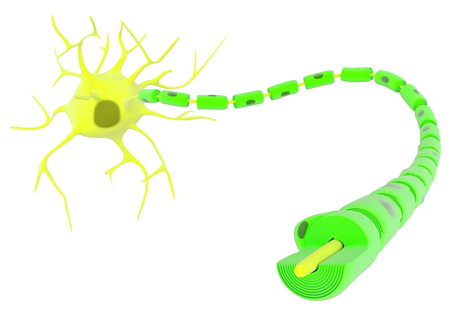
Myelinated neuron anatomy. Illustration of the structure of a nerve cell body (yellow, upper left) and its myelinated axon (right). Myelin (green) is... more
Title
Myelinated Neuron Anatomy
Artist
Science Photo Library
Medium
Photograph
Description
Myelinated neuron anatomy. Illustration of the structure of a nerve cell body (yellow, upper left) and its myelinated axon (right). Myelin (green) is an insulating material that is essential to increase the speed of propagation of the nerve impulse along the axon. Myelin arises from neuroglial cells, shown here with their nuclei (grey). The nerve cell body nucleus (grey) is also shown, along with dendrites branching out from the nerve cell. Dendrites bring signals to the nerve cell, while the axon transmits signals outwards. For this artwork with labels, see C023/8831.
Uploaded
June 28th, 2016
More from Science Photo Library
Comments
There are no comments for Myelinated Neuron Anatomy. Click here to post the first comment.


